Gel electrophoresis is a technique widely used in life science laboratories to separate macromolecules such as DNA, RNA, and proteins. In this technique, molecu
Gel electrophoresis is a technique widely used in life science laboratories to separate macromolecules such as DNA, RNA, and proteins. In this technique, molecules are separated according to size and electrical charge.
How does electrophoresis work?
The gel used in gel electrophoresis is usually made from a material called agarose, which is a jelly-like substance extracted from seaweed. This porous gel could be used to separate macromolecules of many different sizes. The gel is immersed in a salt buffer solution in an electrophoresis chamber. Tris-borate-EDTA (TBE) is commonly used as the buffer. Its main function is to control the pH of the system. The camera has two electrodes - one positive and one negative - at its two ends.
Samplesthatneedtobeanalyzedarethenloadedintotinywellsinthegelwiththehelpofapipette.Oncethechargeisready,anelectriccurrentof50-150Visapplied.Now,thechargedmoleculespresentinthesamplebegintomigratethroughthegeltotheelectrodes.Therateatwhicheachmoleculetravelsthroughthegeliscalleditselectrophoreticmobilityandisdeterminedprimarilybyitsnetchargeandsize.Heavilychargedmoleculesmovefasterthanweaklycharged.Oncetheseparationiscomplete,thegelisstainedwithadyetorevealtheseparationbands.Ethidiumbromideisafluorescentdyecommonlyusedingelelectrophoresis.Thegelissoakedinadilutesolutionofethidiumbromideandthenplacedinanultraviolettransilluminatortovisualizetheseparationbands.Thebandsareexaminedorphotographedimmediatelyforfuturereference,astheywilldiffuseonthegeloveracertainperiodoftime.Gel documentation systems
You should keep in mind that gel electrophoresis is one of the main tools in molecular, cellular and biochemical biology laboratories. In fact, it remains one of the main endorsements and tests requested when publishing important results. This is why researchers require high-resolution and multiple image recognition systems to process gels. From this point of view, the processing of gels requires the highest sensitivity to ensure the quality of the results.
Today, digital development makes available image analysers or gel documentation systems, specifically designed to meet the needs and budget of the laboratory. These provide innovative solutions for both image capture and analysis, and are configured based on the techniques used by each researcher.
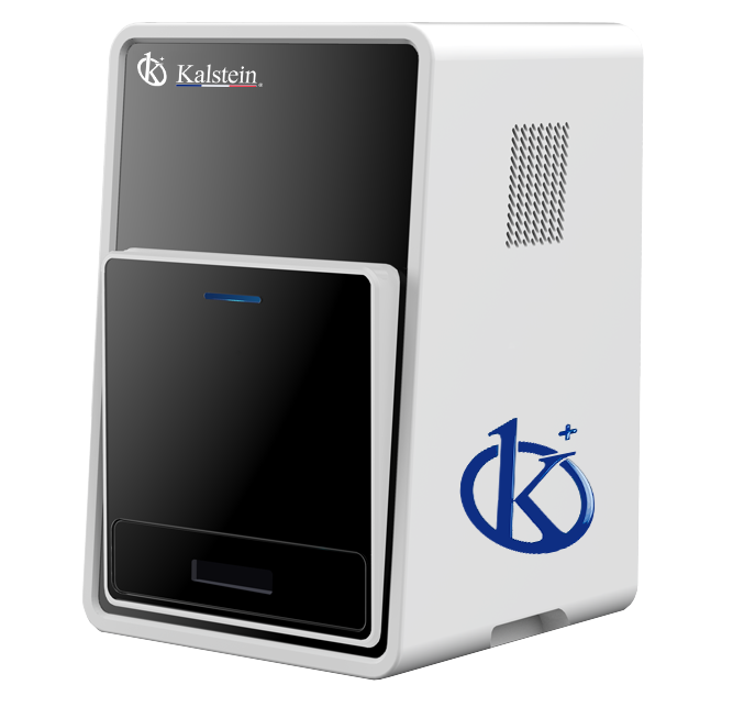
At
Kalsteinwe present you our documentation system for gels whose compact and friendly design allows you to easily capture the image of the gel in high quality. These are systems that have an intuitive user interface, are capable of fast image capture and self-exposure, and come equipped with an exceptional high-resolution camera. Images are easily saved thanks to its software. It has an excellent transilluminator and a blue LED light filter that allows the recovery of nucleic acids directly from the gel, without damage, unlike the old systems. That's why we invite you to take a look at the “Products” menu.
HERE