Angina is temporary chest pain or discomfort. It is clinically defined as a syndrome resulting from myocardial ischaemia (damage or disease to major blood vessels in the heart), caused by decreased blood flow to the heart muscle. It is characterized by discomfort near the breastbone but can be felt anywhere on the body, from the epigastrium, jaws, shoulders, back, or arms that occurs during exercise or stress. Symptoms are often described as tension, tightness, or heaviness and may be accompanied by a sensation of constriction, strangulation, or burning.
Angina may be stable when developing during physical activity or unstable when occurring during periods of rest, with the latter manifestation being more severe. Importantly, angina is not a heart attack but is a sign of cardiac risk, so the patient must eliminate possible factors that cause the disease to progress and symptoms to recur or become severe. Tests that are used to diagnose and confirm angina include electrocardiography, chest x-ray, blood tests, stress testing, echocardiography, computed tomography of the heart, cardiac magnetic resonance imaging, and coronary angiography.
Electrocardiogram as initial diagnostic test for angina
Electrocardiography should be done initially in any patient with suspected angina to detect angina. The study technique allows to determine signs of ischemic heart disease, such as a previous myocardial infarction or alterations of the repolarization, showing the rhythm of functioning of the heart. It should be noted, this resting test is quite common even in patients with severe angina and does not exclude the diagnosis of ischemia. The methodology is quick and painless, sensitive and specific to measure the electrical activity of the heart. Preferably used to identify inducible ischemia in most patients with stable angina.
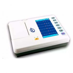
What is an electrocardiogram and how does it work?
The electrocardiograph is an electronic device that captures and expands the electrical activity of the heart through electrodes and the information captured is recorded on an electrocardiogram. The electrocardiograph was described by the Dutch doctor Willem Einthoven in 1903, who was named Nobel Prize in medicine in 1924 for his contributions in the field of health.
This type of medical equipment is part of the bioinstrumentation in biomedical engineering, which is dedicated to record specific biosignals of the human body; and thus, process them to be used by specialists in the area of health, so that the diagnostic process is not invasive.
An electrocardiograph uses electrodes to detect and convert the flow of ions through the heart muscle to electrical current. Because the captured signal is almost imperceptible, a preamplifier is used, which increases the magnitude of the signal to work better with it. Then, the signal is cleaned through filters and a final amplification is carried out for their respective digitization.
There are several types of electrocardiographs including
- Single-channel electrocardiograph: characterized by recording and printing the results of the heart’s electrical activity in a single lead per record.
- Multichannel electrocardiograph: distinguishes itself by having 3.6 or 12 channels and recording each of the 12 leads in a period of 2.5 seconds.
- Print multichannel: Different from the above because they have a computer with recognition patterns that identifies normal and abnormal electrocardiogram signals.
At Kalstein we are MANUFACTURERS and we offer you new electrocardiographic units. If you are interested in knowing our products, the PRICES for BUY or SALE visit our website HERE