Multiple Myeloma is a multifocal plasma cell neoplasm that affects the bone marrow and is associated with the production of a serum and urine monoclonal protein. The cause is progressive unregulated proliferation of plasma cells that accumulate in the bone marrow. These cells secrete immunoglobulin (Ig) excessively; typically: IgG 57%, IgA 21%, IgD 1%, IgM, IgE, only rarely in 18% of cases of light chains alone. Proliferation of multiple myeloma interferes with the normal production of cells in the bone marrow and typically results in anemia. Leukopenia and thrombocytopenia occasionally occur. Another feature is that multiple myeloma cells secrete certain osteoclast-stimulating and osteoblast-inhibiting substances that result in exaggerated destruction of bone tissue with subsequent pathologic fracture, in many cases hypercalcemia.
What is the pathogenesis of Multiple Myeloma?
Pathogenesis includes monoclonal gammopathy of undetermined significance (MGUS) and asymptomatic MM has been determined that translocations that compromise the locus of the immunoglobulin heavy chain on chromosome 14q32, start and maintain the proliferative clone, which is accompanied by other chromosomal alterations and gene dysregulation especially of the D1, D2 or D3 cyclins, reaching a prognostic classification, where the genetic profile plays a role of importance.
What is M-protein?
As mentioned above, a characteristic feature of multiple myeloma is the production of a monoclonal protein (M-protein). The test used to measure the amount of monoclonal protein in blood or urine is Protein Electrophoresis.
This M-protein is synthesized by malignant plasma cells that are immunoglobulin molecules or parts of them. The amount of protein produced and secreted into the serum and urine reflects the state of myeloma in the body at any given time.
What is Protein Electrophoresis? Serum?
Protein electrophoresis is a laboratory test based on the separation of proteins using an electric field. When a sample containing a mixture of different proteins is electrophoretic, the different proteins in the mixture are separated according to their electrical charge.
These fractions (also called “zones” or “regions”) are called albumin, Alpha 1, Alpha 2, Beta (which can be separated into Beta 1 and Beta 2), and Gamma. Polyclonal (normal) immunoglobulins are in the Gamma zone.
Normal serum immunoglobulins are diverse and show slight differences in their structure and electrical charge.
Monoclonal proteins are produced by a clone of plasma cells, so all molecules are identical and have the same electrical charge. That is why a monoclonal protein will migrate like a narrow peak in electrophoresis (the peak appears almost always in the gamma zone, but may sometimes be present in Beta 2 or Beta 1; or even in the Alpha 2 zone, although the latter is uncommon). Serum electrophoresis can be used to search for a monoclonal protein as well as to monitor the amount of monoclonal protein present.
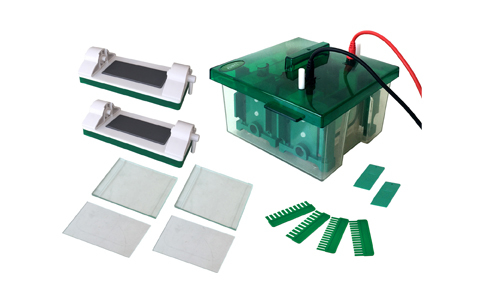
At Kalstein we offer you a high quality system that will help you to determine the Electrophoresis of proteins, so that you obtain in this way much more accurate results and with a high rate of reproducibility. So we invite you to take a look: HERE

