Calcium (Ca) is an alkaline earth metal that fulfills important functions in the body. The metal circulating in the blood is 45% bound to plasma proteins, mainly albumin, and 15% to small anions. The remaining 40% of circulating calcium is in its ionic form. This means that you can measure, on the one hand:
- Total calcium (standard value: 8.5-10.5 mg/dl or 2.12-2.62 mmol/l).
- Ionized calcium (standard: 4.65-5.25 mg/dl or 1.16-1.31 mmol/l).
In calcium deficiency or hypocalcemia, the body lacks the mineral calcium. Doctors speak of calcium deficiency when the total calcium level in blood falls below 2.2 mmol/l. As the name suggests, calcium deficiency means that the body does not receive enough calcium. Adults need about 1,000 mg of calcium a day.
Pregnant and breastfeeding women, as well as growing children and adolescents, need more. The additional need for calcium can be met by dairy products but only if there is no cow’s milk allergy or lactose intolerance. Soy and soy products are also especially rich in calcium, as are green vegetables such as kale, broccoli, or fennel.
What causes hypocalcemia?
- Vitamin D deficiency.
- Calcium metabolism disorders congenital.
- Chronic renal failure.
- Acute pancreatitis.
- Rhabdomyolysis.
- The sepsis.
Hypocalcemia is frequently observed in critically ill patients, with a reported incidence of up to 50%. Also, hypocalcemia is often associated with hypomagnesemia. Neuromuscular irritability is the cardinal feature of hypocalcemia, with carpal-pedal spasm being the classic physical sign that can progress to overt tetany, laryngospasm or tonic-clonic seizure activity.
How is hypocalcemia diagnosed with an electrocardiograph?
The most common electrolyte abnormalities affecting the electrocardiogram include disorders of potassium, calcium, and magnesium. Electrolyte imbalances affect the depolarization and repolarization phases of the cardiac cycle action potential by altering potentials across cardiac myocyte cell membranes. Although each electrolyte and its associated abnormalities can be discussed separately, it is important to remember that combined disorders occur and that there is a dynamic physiologic interaction between these cations.
The main electrocardiographic manifestation of hypocalcemia is QTc interval prolongation. Hypocalcemia prolongs action potential phase 2 with an impact modulated by the rate of change in serum calcium concentration and myocyte calcium channel function. QTc prolongation is associated with early post-repolarizations and triggered dysrhythmias. Torsades de pointes may be triggered by hypocalcemia but is much less common than with hypokalemia or hypomagnesemia.
While ECG conduction abnormalities are common, severe dysrhythmias induced by hypocalcemia, such as heart block and ventricular dysrhythmias, are uncommon. The development of dysrhythmias is often associated with other comorbidities such as structural heart disease, ischemia or in association with drug therapy (e.g. digitalis, catecholamines).
Severe symptoms and life-threatening dysrhythmias require immediate treatment with parenteral calcium salts. In addition, associated electrolyte abnormalities, such as hypomagnesemia, phosphate abnormalities, and acidemia, may need to be corrected. For chronic hypocalcemia, vitamin D and calcium supplements may be given by mouth.
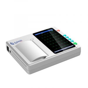
Kalstein electrocardiograph for diagnosis of hypocalcemia
Diagnosis of hypocalcemia can be made rapidly with electrocardiography, although serum calcium measurement is one of the most common diagnostic canons. In particular, the electrocardiograph of the equipment manufacturer Kalstein allows to display on its LCD screen the electrocardiogram as it is being obtained, has a thermal printing system, low noise level and lifetime warranty. For more information on these devices, such as pricing, purchase, or request for a quote, visit HERE