Inflammation of the heart muscle (myocarditis) is a serious disease. Symptoms are often difficult to recognize, making rapid diagnosis difficult. In severe cases, myocarditis may cause heart failure or severe heart rhythm disturbances. It often develops after flu-like infections. Other possible causes include:
- Viral infections such as herpes, measles or coxsackie virus
- Bacterial infections, for example, the pathogens of tonsillitis, scarlet fever, diphtheria, or blood poisoning
- Parasitic infections
- Drugs
- Side effects of radiation therapy
Because of atypical clinical features, the diagnosis of myocarditis is often made only after other potentially life-threatening differential diagnoses have been ruled out. Crucial diagnostic procedures include electrocardiography (the most widespread examination of all), cardiac biomarkers such as troponin T, and cardiac echocardiography and magnetic resonance imaging.
The gold standard is myocardial biopsy, but because it is an invasive procedure, it is not done regularly. Immunohistochemical analysis of myocardial biopsy should be done in a cardiology center that allows an invasive examination option in the cardiac catheterization laboratory.
What are the symptoms of myocarditis?
Viruses or bacteria usually cause inflammation of the heart muscle (infectious myocarditis). Thus, symptoms of such infection usually precede myocarditis. They include a cold and cough, fever, headache, and pain in the limbs.
If these flu-like symptoms are accompanied by increased fatigue and exhaustion, weakness, a decreased ability to recover, or shortness of breath on exertion, they suggest infectious myocarditis. However, there are no clear or definite main symptoms. In fact, these discomforts are usually the only signs at the onset of acute myocarditis. Sometimes symptoms such as loss of appetite and weight and pain radiating from the neck or shoulders are added.
How can myocarditis be diagnosed?
Viral inflammatory processes may be well detected by laboratory, long-term ECG, echocardiography, computed tomography (CT), magnetic resonance imaging (MRI), or catheter-based diagnosis. Thus, systolic or diastolic heart failure, hemodynamic arrhythmias, or valvular system damage can be diagnosed qualitatively and quantitatively, and dilated, restrictive, or hypertrophic cardiomyopathies can be distinguished.
Cardiac MRI should be done in symptomatic patients with clinical evidence of myocarditis. In patients with clinically suspected myocarditis, acute changes on infarction-type ECG, positive troponin T/I, elevation of NT-pro-BNP, and evidence of positive edema or early enhancement are nonspecific indicators of virus- or inflammatory cell-associated myocardial injury.
However, responsible toxic, infiltrative, or infectious-inflammatory processes at the cellular level are not detected or are insufficiently detected by noninvasive clinical diagnoses, including MRI. On the other hand, in ECG studies, certain changes can be detected that denote the presence of a condition in the heart and that merits more detailed analysis. In this sense, the ECG may have the following changes:
- QRS complex extended
- Left bundle branch block
- ST, segment, and T wave changes
- Bradycardic or tachycardic arrhythmias
- Signs of infarction
- Low voltage and P wave changes
- Signs of concomitant pericarditis
Because this test is based on measuring the electrical activity of the heart muscle (electrocardiography, ECG), it can detect these changes in heart activity as they result from inflammation of the heart muscle. Typical symptoms are rapid heartbeat (palpitations) and extra beats (extrasystoles). Cardiac arrhythmias are also possible. Because deviations usually occur only temporarily, a long-term measurement of cardiac activity (long-lasting ECG) in addition to the usual short-lived resting ECG is advisable.
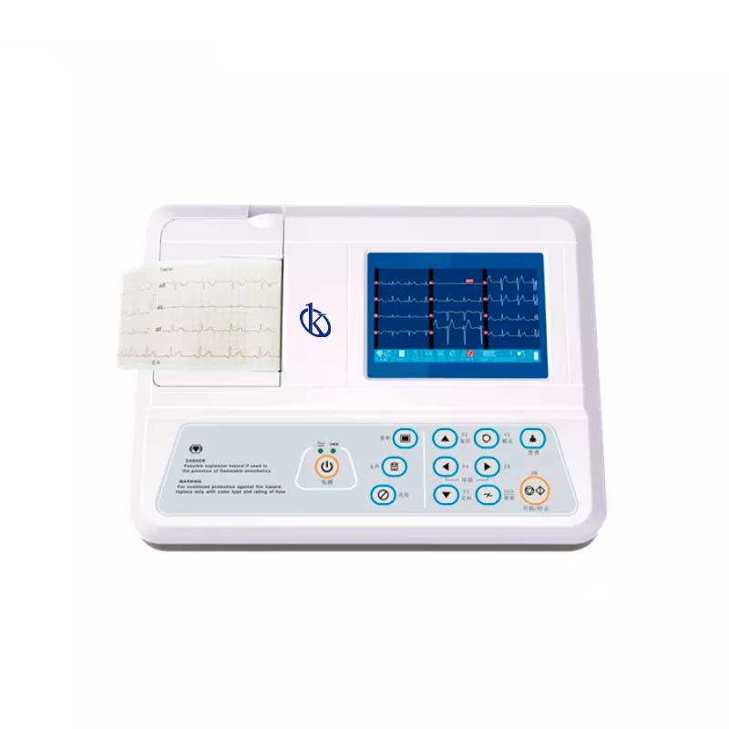
Why use Kalstein’s electrocardiograph to diagnose myocarditis?
Kalstein’s electrocardiograph is designed and constructed by the highest quality standards, which guarantee the measurements that support the diagnosis made. In this sense, the electrocardiographs produced by this manufacturer are characterized by deriving up to twelve signals simultaneously, automatic correction of the drift of the baseline, also has a low noise level. Pricing, purchase and quotes for these devices can be found at HERE or HERE

